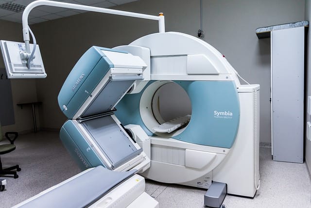
Neuroimaging (Brain Imaging) has revolutionized our understanding of the human brain, providing critical insights into its structure and function. By employing various imaging techniques, researchers can visualize and analyze brain activity, revealing the intricate workings of neural circuits that underpin behavior, cognition, and emotion. This post offers an overview of Brain Imaging, detailing its significance in psychology and psychiatry and its applications in diagnosing and treating various mental health conditions.
The landscape of Brain Imaging is vast, encompassing techniques such as functional magnetic resonance imaging (fMRI), electroencephalography (EEG), and diffusion tensor imaging (DTI). Each method serves unique purposes and contributes to our understanding of different aspects of brain function. For instance, functional Brain Imaging allows scientists to observe brain activity in real time, while structural Brain Imaging provides detailed images of brain anatomy.
Understanding Brain Imaging is crucial for both researchers and clinicians. The insights gained from these techniques not only advance scientific knowledge but also inform treatment strategies for conditions such as schizophrenia, ADHD, and depression. In this blog, we will explore the various types of Brain Imaging , their applications in clinical settings, and the future directions of this exciting field.
The Evolution of Neuroimaging
Historical Perspective
Brain Imaging has undergone a remarkable evolution since its inception. The journey began in the late 19th century with the advent of early imaging techniques like X-rays. However, it was not until the development of MRI technology in the late 20th century that significant breakthroughs occurred. MRI allowed for non-invasive imaging of soft tissues, paving the way for more detailed studies of brain structure and function.
Milestones in Neuroimaging
Key milestones in the history of Brain Imaging include the first functional MRI scan in the early 1990s, which enabled researchers to visualize brain activity in real-time. This was followed by advancements in PET scans and DTI, further expanding the toolkit available to neuroscientists. Today, Brain Imaging plays a central role in both research and clinical practice, providing invaluable insights into neurological disorders and cognitive processes.
Types of Neuroimaging
Functional Neuroimaging
Functional Brain Imaging , particularly fMRI, has become a cornerstone in understanding brain activity. By measuring changes in blood flow, researchers can infer which areas of the brain are involved in specific tasks or processes. For more information, visit Frontiers in Neuroimaging.
Key Applications of Functional Neuroimaging
Cognitive Research: Functional Brain Imaging is essential for understanding how different cognitive processes are represented in the brain. Studies have demonstrated the involvement of specific brain regions in memory, attention, and decision-making.
Clinical Applications: In clinical settings, fMRI can assist in pre-surgical planning by identifying critical brain areas responsible for essential functions, such as language and motor skills. This ensures that surgeons minimize damage to these areas during procedures.
Structural Neuroimaging
Structural Brain Imaging techniques, such as MRI and CT scans, provide detailed images of brain anatomy. These methods are essential for diagnosing conditions like tumors or brain injuries. Structural brain imaging is particularly important in clinical Brain Imaging for understanding various psychiatric disorders.
Importance of Structural Neuroimaging
Diagnosing Brain Disorders: Structural Brain Imaging plays a critical role in diagnosing brain disorders, including tumors, neurodegenerative diseases, and traumatic brain injuries.
Understanding Brain Development: Researchers use structural Brain Imaging to study brain development across the lifespan, helping to identify atypical patterns associated with developmental disorders.
Diffusion Tensor Imaging (DTI)
DTI is a specialized type of MRI that focuses on the diffusion of water molecules in brain tissue, allowing for the visualization of white matter tracts. This technique has significant applications in research related to neuroimaging in schizophrenia and other neurological conditions.
Applications of DTI
White Matter Integrity: DTI is instrumental in assessing white matter integrity, which is crucial for understanding various neuropsychiatric conditions, including schizophrenia and major depressive disorder.
Understanding Connectivity: DTI helps researchers map brain connectivity, revealing how different brain regions communicate with one another. This information is vital for understanding the neural basis of cognitive functions.
Applications of Neuroimaging
Neuroimaging in Psychiatry
Brain Imaging has transformative potential in psychiatric research and practice. By employing brain imaging techniques, researchers can identify biomarkers for mental health disorders, leading to earlier diagnoses and tailored treatment approaches. For insights into the intersection of Brain Imaging and psychiatry, explore Psychiatry Research: Neuroimaging.
Key Contributions to Psychiatry
Identifying Biomarkers: Brain Imaging studies have identified brain-based biomarkers that correlate with symptoms of various psychiatric disorders, aiding in early diagnosis and personalized treatment plans.
Understanding Treatment Effects: By examining brain changes in response to therapeutic interventions, Brain Imaging can help clinicians optimize treatment strategies and evaluate their efficacy.
ADHD and Neuroimaging
Studies focusing on ADHD Brain Imaging have uncovered differences in brain structure and function among individuals with ADHD compared to neurotypical individuals. This research helps to elucidate the neurological basis of the disorder and informs potential treatment strategies.
Insights from ADHD Neuroimaging
Structural Differences: Research has shown that individuals with ADHD may have structural differences in regions related to attention and impulse control, such as the prefrontal cortex and basal ganglia.
Functional Differences: Functional neuroimaging studies reveal altered activation patterns in the brains of individuals with ADHD during tasks requiring sustained attention, providing insights into the cognitive deficits associated with the disorder.
Multimodal Neuroimaging
Combining different Brain Imaging techniques, known as multimodal Brain Imaging, allows for a comprehensive understanding of brain function. By integrating structural and functional data, researchers can gain deeper insights into the neural correlates of behavior and cognitive processes. For more information, visit J Neuroimaging.
Benefits of Multimodal Approaches
Holistic Understanding: Multimodal Brain Imaging provides a holistic view of brain function, revealing how structure and function interact to support cognition and behavior.
Improving Diagnostic Accuracy: By combining data from multiple imaging modalities, researchers can improve diagnostic accuracy and better understand the Brain Imaging underpinnings of psychiatric disorders.
Neuroimaging Research and Journals
Numerous journals publish cutting-edge research in the field of Brain Imaging , including the J Neuroimaging and Biological Psychiatry: Cognitive Neuroscience and Neuroimaging. These journals provide a platform for disseminating important findings that contribute to our understanding of the brain.
Key Journals in Neuroimaging
J Neuroimaging: This journal focuses on clinical Brain Imaging and publishes research on various imaging techniques and their applications in clinical practice.
Frontiers in Neuroimaging: A leading journal that covers a wide range of Brain Imaging topics, including methodological advancements and clinical applications.
Emerging Trends in Neuroimaging
Artificial Intelligence and Neuroimaging
The integration of artificial intelligence (AI) in Brain Imaging is a rapidly growing field. Machine learning algorithms are being developed to analyze complex Brain Imaging data, enabling researchers to identify patterns and biomarkers that may not be apparent through traditional analysis methods.
Applications of AI in Neuroimaging
Automated Analysis: AI can automate the analysis of Brain Imaging data, reducing the time and effort required for manual interpretation.
Predictive Modeling: Machine learning models can predict outcomes based on Brain Imaging data, aiding in early diagnosis and treatment planning.
Neuroimaging in Neurodevelopmental Disorders
Research on neuroimaging in neurodevelopmental disorders, such as autism spectrum disorder (ASD), is expanding. Neuroimaging studies are uncovering the neural correlates of ASD, shedding light on the underlying mechanisms of the disorder.
Insights from Neuroimaging in ASD
Brain Connectivity Patterns: Studies have shown that individuals with ASD may exhibit atypical brain connectivity patterns, impacting social communication and behavior.
Identifying Subtypes: Brain Imaging can help identify subtypes within the ASD population, allowing for more tailored interventions based on individual profiles.
Challenges and Limitations in Neuroimaging
Technical Limitations
Despite its advancements, Brain Imaging faces several technical challenges. Limitations in spatial and temporal resolution can affect the accuracy of findings, particularly in studying dynamic brain processes.
Ethical Considerations
Brain Imaging research also raises ethical considerations, particularly regarding data privacy and informed consent. Researchers must navigate these challenges to ensure ethical standards are upheld in their studies.
Future Directions in Neuroimaging
Advancements in Imaging Techniques
The future of Brain Imaging holds promise with advancements in imaging techniques. New modalities and improved technologies are being developed to enhance the resolution and accuracy of Brain Imaging studies.
Clinical Applications and Personalized Medicine
As our understanding of the brain improves, the clinical applications of neuroimaging will continue to expand. Personalized medicine approaches, informed by Brain Imaging data, will likely play a crucial role in treating mental health conditions.
Conclusion
In conclusion, Brain Imaging is a vital field that continues to advance our understanding of the human brain. With various imaging techniques available, researchers are uncovering the complex relationships between brain structure and function, leading to significant advancements in the diagnosis and treatment of mental health conditions.
As we delve deeper into the world of Brain Imaging , the implications for psychology and psychiatry become increasingly profound. Engaging with these technologies opens new avenues for understanding and addressing neurological and psychiatric disorders, ultimately enhancing the quality of care for individuals affected by these conditions. The future of neuroimaging is bright, promising to unveil even more about the intricate workings of the brain and its impact on behavior, cognition, and emotional well-being.


