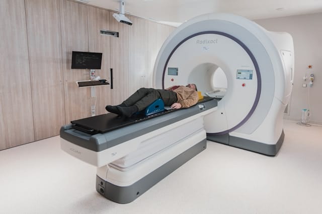
Positron Emission Tomography, commonly known as PET, is a powerful imaging technique that plays a crucial role in modern medicine. This technology allows for the visualization of metabolic processes in the body, providing insights that are critical for diagnosing various diseases, particularly cancer. In this blog post, we will explore ten essential insights into positron emission tomography, from its underlying principles to its clinical applications and future directions.
Introduction to Positron Emission Tomography
At its core, positron emission tomography is a non-invasive imaging technique that allows medical professionals to observe metabolic activity within the body. By employing radioactive tracers that emit positrons, PET scans can detect the gamma rays produced when positrons interact with electrons in the body. This process helps create detailed images of organs and tissues, enabling healthcare providers to make informed decisions regarding diagnosis and treatment.
The development of positron emission tomography has revolutionized medical imaging, offering a unique perspective on how diseases affect the body at the cellular level. Unlike traditional imaging methods such as X-rays or MRIs, which primarily show anatomical structures, PET provides functional information that can reveal underlying health issues long before they manifest as structural abnormalities.
In this article, we will delve into ten essential insights about positron emission tomography, covering its history, technology, clinical applications, and its significance in advancing medical research. Whether you’re a medical professional, a student, or simply curious about this remarkable technology, this guide will provide a comprehensive understanding of PET.
The History of Positron Emission Tomography
The journey of positron emission tomography began in the mid-20th century, driven by advancements in nuclear medicine and imaging technologies. The first concepts for PET emerged from the work of physicists and engineers who sought to combine the principles of radioactive decay with imaging techniques.
The Birth of PET
The first practical positron emission tomography scanner was developed in the early 1970s by Dr. Michael E. Phelps and his colleagues at the University of California, Los Angeles (UCLA). Their pioneering work laid the groundwork for the modern PET scanner, which was capable of producing 3D images of the body’s metabolic activity. This breakthrough was made possible by the discovery of fluorodeoxyglucose (FDG), a radioactive glucose analog, which could be used as a tracer to visualize cancerous tissues.
The introduction of PET technology offered a new dimension to medical imaging. Before PET, imaging modalities primarily focused on structural imaging, which did not account for the biological processes that occur within the body. The ability to visualize metabolic activity marked a significant shift in diagnostic capabilities.
Advancements in Technology
Since its inception, positron emission tomography has undergone significant technological advancements. Early PET scanners were bulky and limited in their capabilities. However, innovations in detector technology, image reconstruction algorithms, and computer processing power have dramatically improved the quality and speed of PET imaging. Today’s scanners can produce high-resolution images with greater sensitivity, allowing for the detection of smaller lesions and improved diagnostic accuracy.
One of the significant advancements in PET technology has been the introduction of Time-of-Flight (TOF) PET imaging. TOF PET uses the time difference between the detection of two gamma rays emitted from a positron-electron annihilation event to improve image quality. This technology reduces image noise and increases the sensitivity of the scanner, allowing for more precise detection of tumors and other abnormalities.
How Positron Emission Tomography Works
The fundamental principle behind positron emission tomography involves the use of radioactive tracers that emit positrons during their decay process. This section will break down the key components and steps involved in a typical PET scan.
Radioactive Tracers
At the heart of positron emission tomography is the use of radioactive tracers. These tracers are usually molecules that have been labeled with a radioactive isotope, such as fluorine-18 or carbon-11. When administered to a patient, the tracer accumulates in specific tissues or organs based on their metabolic activity. For example, FDG is preferentially taken up by cancer cells, which metabolize glucose at a higher rate than normal cells.
The selection of a tracer is critical, as it determines the type of biological processes that can be visualized. For instance, specific tracers are developed to target various types of tumors, neurodegenerative diseases, and cardiovascular conditions. The versatility of tracers expands the applications of PET beyond oncology to other areas of medicine.
The Imaging Process
The imaging process begins with the intravenous injection of the radioactive tracer. After a brief waiting period, during which the tracer distributes throughout the body, the patient is placed in the PET scanner. The scanner detects the gamma rays emitted when positrons from the tracer collide with electrons in the body, resulting in annihilation events that produce pairs of gamma rays.
The scanner collects data from these gamma rays, which are then processed using sophisticated algorithms to reconstruct detailed images of the body. These images illustrate areas of increased metabolic activity, providing valuable information about the patient’s health status.
Image Reconstruction Techniques
The data collected during a PET scan undergoes a complex image reconstruction process. This involves several steps, including:
- Data Acquisition: The scanner collects raw data from the detected gamma rays.
- Correction Algorithms: Various corrections are applied to account for factors such as scattering and attenuation of gamma rays. These corrections improve the accuracy of the images.
- Reconstruction Algorithms: Advanced algorithms, such as Filtered Back Projection (FBP) or Iterative Reconstruction (IR), are used to create a visual representation of the distribution of the tracer in the body. Iterative reconstruction methods are particularly advantageous as they provide better image quality and reduce noise.
The result is a set of high-resolution images that can be analyzed to assess metabolic activity in different tissues.
Clinical Applications of Positron Emission Tomography
Positron emission tomography has a wide range of clinical applications, making it an invaluable tool in the diagnosis and management of various medical conditions. Below are some of the key areas where PET is commonly utilized.
Cancer Diagnosis and Staging
One of the primary uses of positron emission tomography is in the detection and staging of cancer. PET scans can reveal the metabolic activity of tumors, helping oncologists determine the presence and extent of cancerous lesions. This information is critical for planning treatment strategies, monitoring response to therapy, and assessing recurrence after treatment.
PET scans are particularly valuable in distinguishing between benign and malignant tumors. For example, a tumor may show increased FDG uptake, indicating higher metabolic activity consistent with malignancy. Conversely, benign lesions may demonstrate lower metabolic activity. This capability enhances the accuracy of diagnosis and informs treatment decisions.
Neurological Disorders
PET is also instrumental in the evaluation of neurological disorders. Conditions such as Alzheimer’s disease, Parkinson’s disease, and epilepsy can be assessed using PET scans, as they allow for the visualization of brain metabolism and function. For instance, a PET scan can detect decreased glucose metabolism in specific regions of the brain associated with Alzheimer’s disease, aiding in early diagnosis.
In addition to diagnosing neurological conditions, PET is used to evaluate brain tumors and assess the effectiveness of treatments. Monitoring changes in metabolic activity can provide insights into tumor response to therapy and help guide subsequent treatment decisions.
Cardiovascular Imaging
In the realm of cardiovascular medicine, positron emission tomography is used to assess myocardial perfusion and viability. PET scans can identify areas of the heart muscle that may be ischemic or scarred due to previous heart attacks. This information helps guide treatment decisions, including the need for revascularization procedures.
PET can also be used to evaluate coronary artery disease by assessing blood flow to the heart muscle during stress and rest. By visualizing areas of reduced blood flow, healthcare providers can identify blockages and determine the best course of action for the patient.
Advantages of Positron Emission Tomography
Positron emission tomography offers several advantages over traditional imaging modalities, making it a preferred choice in many clinical settings.
Functional Imaging
Unlike conventional imaging techniques that primarily provide anatomical information, PET offers functional insights into metabolic processes. This capability allows for earlier detection of diseases, as changes in metabolic activity often occur before structural changes are visible on other imaging tests.
For example, in cancer diagnosis, a PET scan may reveal increased glucose metabolism in a tumor even when the tumor size is not sufficient to be detected on a CT scan. This early detection is crucial for initiating timely treatment, which can significantly improve patient outcomes.
High Sensitivity and Specificity
Positron emission tomography is known for its high sensitivity and specificity in detecting various diseases. The ability to visualize metabolic activity means that PET can identify abnormalities that might be missed by other imaging techniques, enhancing diagnostic accuracy.
This high sensitivity is particularly important in oncology, where the early detection of small tumors can influence treatment decisions and improve survival rates. PET’s specificity also helps distinguish between different types of lesions, allowing for more targeted therapies.
Non-Invasiveness
PET scans are non-invasive and generally well-tolerated by patients. The procedure typically takes less than an hour, and the radiation exposure from the radioactive tracers is relatively low, making it a safe option for many patients.
The non-invasive nature of PET imaging means that it can be repeated multiple times throughout a patient’s treatment journey, allowing for ongoing monitoring of disease progression or response to therapy. This capability is invaluable in managing chronic conditions such as cancer and neurological disorders.
Limitations of Positron Emission Tomography
Despite its many advantages, positron emission tomography also has certain limitations that should be considered.
Limited Availability
One of the primary challenges associated with positron emission tomography is the limited availability of PET scanners and radiopharmaceuticals. The production of radioactive tracers is complex and requires specialized facilities, leading to longer wait times for patients in some areas.
The infrastructure required to support PET imaging, including radiopharmaceutical production and specialized imaging centers, can be expensive. This limited availability can create disparities in access to PET scans, particularly in rural or underserved areas.
Radiation Exposure
While the radiation exposure from a PET scan is relatively low, it is still a consideration, especially for patients requiring multiple scans or for pediatric patients. Careful risk-benefit analysis is essential to minimize unnecessary radiation exposure.
Healthcare providers must weigh the benefits of obtaining critical diagnostic information against the potential risks associated with radiation exposure. In some cases, alternative imaging modalities may be considered if the risks are deemed too high.
False Positives and Negatives
PET scans can sometimes produce false positive or negative results. For example, inflammation or infection may lead to increased metabolic activity that mimics cancer, resulting in a false positive diagnosis. Conversely, small tumors may not exhibit sufficient metabolic activity to be detected, leading to false negatives. Careful interpretation of PET images in conjunction with other diagnostic information is crucial to avoid misdiagnosis.
To mitigate these issues, clinicians often use PET scans in combination with other imaging techniques, such as CT or MRI, to provide a more comprehensive view of the patient’s condition. This multimodal approach enhances diagnostic accuracy and reduces the likelihood of misinterpretation.
The Future of Positron Emission Tomography
As technology continues to advance, the future of positron emission tomography looks promising. Several trends and developments are poised to enhance the capabilities of PET in medical imaging.
Novel Radiotracers
Research is ongoing to develop new radiotracers that target specific biological processes. These novel tracers could improve the specificity of PET scans, allowing for the detection of a wider range of diseases. For instance, radiotracers that target specific cancer cell receptors or biomarkers could enable more precise imaging and personalized treatment approaches.
The development of tracers that can differentiate between various tumor types and their metabolic profiles is particularly exciting. This advancement could pave the way for tailored therapies that are more effective and have fewer side effects.
Hybrid Imaging Technologies
The integration of positron emission tomography with other imaging modalities, such as computed tomography (CT) or magnetic resonance imaging (MRI), has gained popularity. Hybrid imaging systems, such as PET/CT or PET/MRI, combine the functional information from PET with the anatomical detail from CT or MRI, providing a comprehensive view of the patient’s condition. This approach enhances diagnostic accuracy and allows for more effective treatment planning.
Hybrid imaging can also improve the localization of lesions, making it easier for surgeons to plan interventions. The combination of functional and anatomical information can lead to better outcomes in various clinical settings, from oncology to neurology.
Machine Learning and Artificial Intelligence
The application of machine learning and artificial intelligence in analyzing PET images holds great potential. These technologies can improve image reconstruction, enhance diagnostic accuracy, and aid in the interpretation of complex PET scans. By leveraging large datasets and advanced algorithms, AI can assist radiologists in making more informed decisions and improving patient outcomes.
AI-driven tools can help identify patterns in PET imaging data that may not be immediately apparent to human observers. This capability can enhance the early detection of diseases, improve treatment planning, and provide more personalized patient care.
Conclusion: The Impact of Positron Emission Tomography
In conclusion, positron emission tomography is a transformative imaging technique that has revolutionized the field of medicine. From its historical development to its wide-ranging clinical applications, PET has become an indispensable tool for diagnosing and managing various medical conditions. While challenges remain, ongoing research and technological advancements continue to enhance the capabilities of positron emission tomography, paving the way for improved patient care.
By understanding the principles and applications of positron emission tomography, healthcare professionals and patients alike can appreciate the significance of this remarkable technology in advancing medical diagnostics and treatment strategies. As we look to the future, the continued integration of innovative technologies and research will undoubtedly drive further improvements in PET, ultimately leading to better outcomes for patients worldwide.


